Following on from our article covering routine corrective/preventative hoof trimming, we’ll now look at treating specific foot issues beyond the routine trim. In the part 1 article, we covered Steps 1-3 of the Dutch five-step method. In this part 2 article, we’ll cover Steps 4 and 5 of this method, but firstly we’ll go over some common lesions, their appearance, and how they can occur.
Common lesions
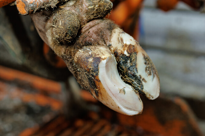
Solar haemorrhage (bruising) or ulcer
These are variations of similar conditions, with bruising often leading to ulcers if not treated.
Risk factors include: long toes (increasing the weight placed on the rear of the sole, where bruising and ulcers are most likely to occur), long standing times at milking, the period around calving (ligaments loosen at calving, causing the bones in the foot to press more against the rear of the sole), heifers (young cattle naturally have thinner soles and less fat in the fat pad lying beneath the bones of the foot), and thin or old cows (their fat pad in the foot is reduced and can be more fibrous).
Bruising looks like reddish/dark brown areas on the sole, generally on the inner rear two thirds of the claws. The feet are often tender, and this can affect more than one foot. If bruising is all that is identified when the feet are lifted, a good place to start for treatment is a course of NSAIDs (non-steroidal anti-inflammatory drugs) such as Ketomax or Metacam and reduced walking distance for a few days.
Ulcers appear in the same locations as bruises but are far more severe. They are not caused by stones piercing the hoof, but form by pressure from the underlying tissue eroding away the hoof sole. This exposes sensitive tissue and leads to a very painful lesion, generally located on the middle of the rear two thirds of the sole. When trimming to treat an ulcer, any underrun sole should be removed. The mainstay of treatment is stopping weight bearing on the affected area by blocking the other claw, plus providing pain relief.
As well as reducing risk factors, another way of trying to avoid both of these lesions is by 'dishing out' the claws, as described in Step 3 of the Dutch five-step method (covered in the previous article).
White line disease
The 'white line' is the border between the sole of the hoof and the wall of the hoof. It can be seen as a white line on the sole that follows parallel to the hoof wall. As it's a join between the two surfaces, it's a weak point and is prone to getting stones and debris lodged in it.
Risk factors include: thin soles, soft horn (due to wet conditions, biotin deficiency, etc.), long walking distances, stoney walkways, sideways forces on the feet (hard turns at excessive speeds, etc.), slippery yards, poor cow flow (narrow passageways or other facility design, etc.), and overcrowding.
This type of lesion can present in a range of severities, varying from light damage or small shallow stones lodged in the line, all the way to deep penetration that can cause abscesses to form under the sole or back up into the heel bulb.
Treatment involves trimming the hoof and removing the underrun sole up to the point of attachment, without causing any bleeding. This is covered in Step 4 of the Dutch five-step method below.
Hoof wall crack
A vertical crack found along the inner wall of the claw. These cracks generally start from damage to the skin in between the claws, leading to interrupted growth and a painful crack forming along the wall of the hoof. They're easy to miss when examining a foot unless you are opening up the toes and carefully scraping away the dirt lodged across the inner walls.
Treatment involves cleaning out the crack and removing lodged debris, then placing a block on the opposite claw to reduce weight bearing on the affected claw while it heals.
Foot rot
An infection of the skin around the foot caused by wet/moist conditions and trapped stones irritating or breaking the skin between the claws. This allows bacteria present in the environment to enter the tissue and cause a very painful infection.
Onset of this condition is rapid and requires antibiotics and NSAIDs (Ketomax or Metacam) to treat. If the stone is still present, it will need to be removed from between the claws.
Care must be taken not to assume a swollen foot is automatically foot rot, without lifting the foot and examining the claws for other lesions. This is often a great example of why even when the answer to lameness seems obvious, the Dutch five-step method should still be followed so as not to miss any other conditions.
Digital dermatitis
This presents as a red/grey round lesion on the skin above the back of the hoof, usually between the heel bulbs. It's very sensitive to touch or even water. It can also appear wart-like in later stages of the disease.
Although it's not as commonly seen here in New Zealand compared to other parts of the world, it's still important to keep an eye out for this infection. It is contagious and can quickly spread between cows in your herd. If this lesion is suspected, it's important to get in contact with your vet to discuss the best course of action.
The final steps of the Dutch five-step method
For a preventative or corrective trim, Step 3 of this method (as described in the previous article) may be as far as you need to go.
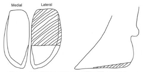
Step 4
Step 4 covers treating claw horn lesions such as solar bruising, ulcers or white line disease. It involves trimming the rear two thirds of the lateral claw. This will lead to increased weight bearing by the medial claw and take pressure off the affected claw, although it is important to preserve the front third of the sole and the inner wall of the hoof as weight bearing surfaces.
Any lesions and surrounding areas should be opened out carefully, without causing any bleeding. Any underrun sole (solar horn that has a pocket under it from an abscess, etc.) should be removed. If it has created an abscess or lead to an infection in the heel bulb, it's important to start these cows on a course of NSAIDs (Ketomax or Metacam) and antibiotics.
Step 5
Step 5 is checking the skin of the foot for infections such as foot rot and digital dermatitis.
After this, if a hoof lesion was identified and treated in Step 4, a block may need to be applied to the opposite claw to take weight off the affected claw. Alongside prompt treatment and pain relief, this is a key aspect of reducing the effect of lameness on cow comfort and production. If in doubt about whether it is required or not, it's always better to improve the comfort of the cow while she recovers. This healing can often take 4-6 weeks depending on the severerity of the lesion.
Source: Bovine Surgery & Lameness Third Edition, by A. David Weaver, Owen Atkinson, Guy St. Jean and Adrian Steiner